Journal of Experimental Botany 63: 5337-5350 (2012)
Embryo growth, testa permeability, and endosperm weakening are major targets for the environmentally regulated inhibition of Lepidium sativum seed germination by myrigalone A [W][OA]
University of Freiburg, Faculty of Biology, Institute for Biology II, Botany / Plant Physiology, Schänzlestr. 1, D-79104 Freiburg, German (AV, KG, KO, DJ, GLM)
School of Biological Sciences, Plant Molecular Science, Royal Holloway, University of London, Egham, Surrey TW20 0EX, Uk (AV, KG, GLM);
Web: 'The Seed Biology Place' - www.seedbiology.eu
Department of Plant Physiology, Warsaw University of Life Sciences-SGGW, 15 Nowoursynowska 159, 02-776, Warsaw, Poland (KO)
Laboratory of Growth Regulators, Faculty of Science, Palacky University and Institute of Experimental Botany AS CR, v.v.i., Šlechtitelu 11, CZ-783 71, Olomouc, Czech Republic (DT, VT, TU, MS)
Centre of the Region Haná for Biotechnological and Agricultural Research, Faculty of Science, Palacky University, Šlechtitelu 11, CZ-783 71, Olomouc, Czech Republic (MS)
Received March 10 2012; Revised June 11 2012; Accepted June 15 2012
Advance Acess publication July 21 2012
DOI 10.1093/jxb/ers197
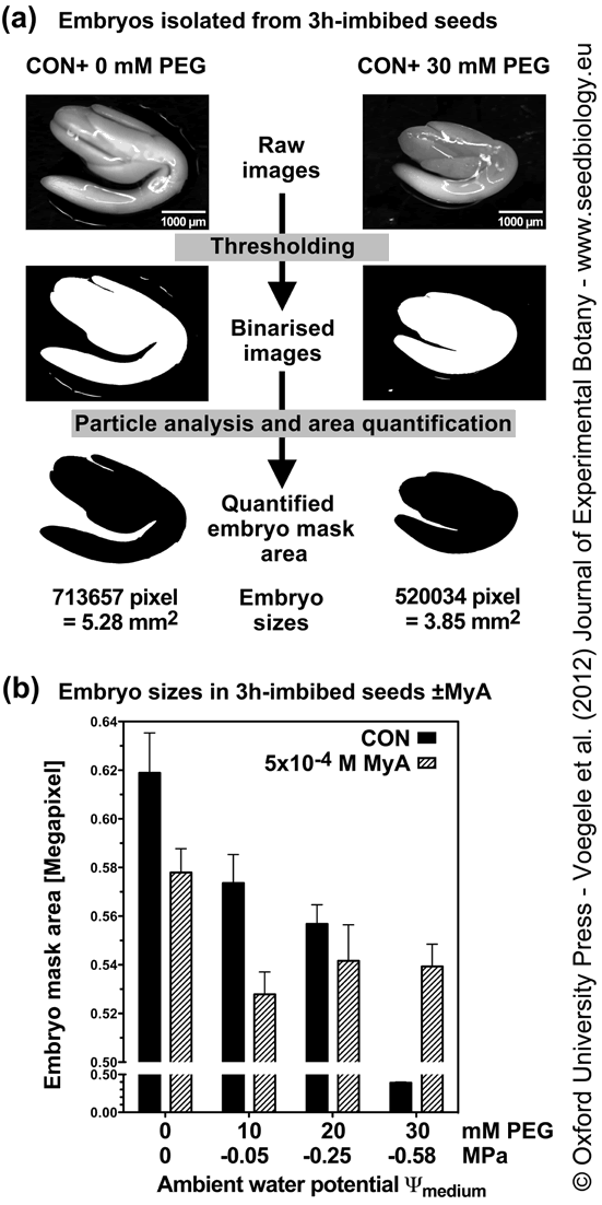
Figure 2. A computer-based image analysis method to quantify Lepidium sativum embryo sizes.
(a) Workflow illustrating quantification of embryo sizes. After seed incubation for a specified time and condition, embryos were carefully extracted and photographed. Raw images were thresholded and image particle analysis was used to create embryo masks. The embryo mask areas were quantified using the Fiji distribution of the image analysis software ImageJ as described in detail in Methods. Using this workflow 1 pixel of the embryo mask area equals 7.4 µm2 real embryo area. Example raw images and mask areas are presented for embryos extracted from 3h-imbibed seeds without or with 30 mM PEG added; pixel- and mm2-values correspond to the presented embryo mask areas.
(b) The effect of decreasing ambient water potential (Ψmedium) on embryo sizes from seeds incubated for 3h in the light without (CON) and with 5x10⁻⁴ M MyA (MyA) and the osmoticum PEG, as indicated.
Mean values ± SE calculated from ≥ 50 extracted embryos are presented.
| Article in PDF format (1.5 MB) Supplementary data file (2 MB) |
|
|
|
The Seed Biology Place |
Webdesign Gerhard Leubner 2000 |