Proteomics 10: 406-416 (2010)
Proteomics reveal tissue-specific features of the cress (Lepidium sativum L.) endosperm cap proteome and its hormone-induced changes during seed germination [W]
Centre National de la Recherche Scientifique-Université Claude Bernard Lyon-Institut National des Sciences Appliquées-Bayer CropScience Joint Laboratory (UMR 5240), Bayer CropScience, Lyon, France (C.J., D.J.)
Centre d'Analyse Protéomique de Marseille, Institut Fédératif de Recherche Jean Roche, Marseille, France (M.B.)
Received July 29, 2009; revised November 3, 2009; accepted November 5, 2009
DOI 10.1002/pmic.200900548
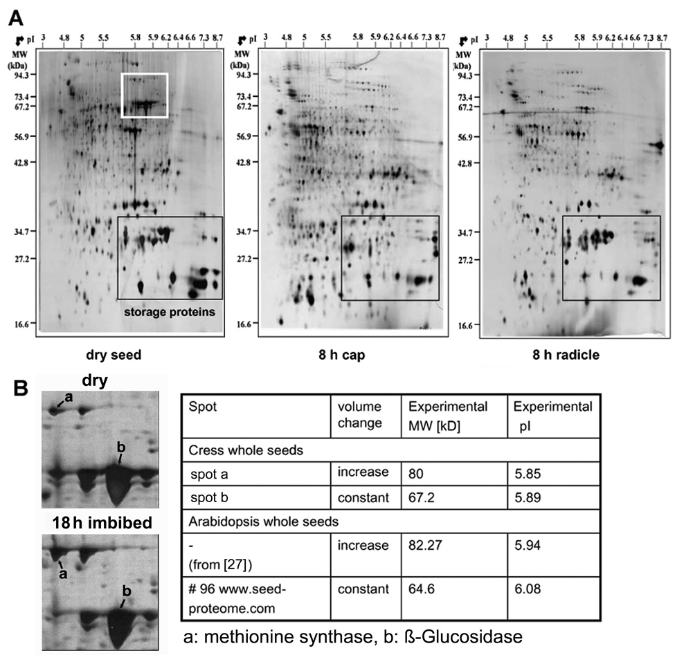
Figure 2. The proteomes of radicle, endosperm cap, and whole cress seeds differ.
(A) Comparison between representative silver-stained 2-D gels of whole dry cress seeds, 8 h imbibed cress radicles and 8 h imbibed cress endosperm caps. Note that the storage proteins (marked by a black square) are most abundant in the whole seed, followed by the radicle. The cap displays a low abundance of storage proteins.
(B) Many cress proteins can already be identified by the similarity of their migration and their volume changes during germination in whole seed extracts to their Arabidopsis orthologous gene products. The section of the dry seed - 2-D gel marked with a white square in (A) is enlarged and compared with the corresponding section of a 2-D gel with extracts of whole cress seeds at 18 h after imbibition (‘‘18 h imbibed’’). The experimental MW and pI of the two spots marked a and b are given in the table to the right of the gel sections and compared with the corresponding Arabidopsis spots. The values for Arabidopsis are taken from [27] and www.seedproteome. com, respectively.
| Article in PDF format (365 KB) Supplemental data file (5 MB) |
|
|
|
The Seed Biology Place |
Webdesign Gerhard Leubner 2000 |